Fotis A. Baltoumas, Ph.D.
  • Home
  • C.V.
  • Research
    • Research Interests
    • Publications
    • Scientific Conferences
    • Theses & Book Chapters
  • Resources
    • Online Tools
    • Other Resources
    • Media Gallery
  • Other
    • Web Development
    • Mystery Magazine
  • Links

Media Gallery


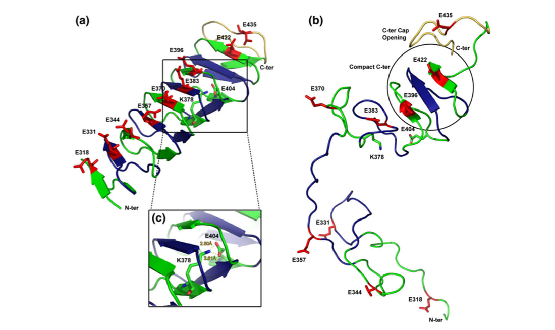
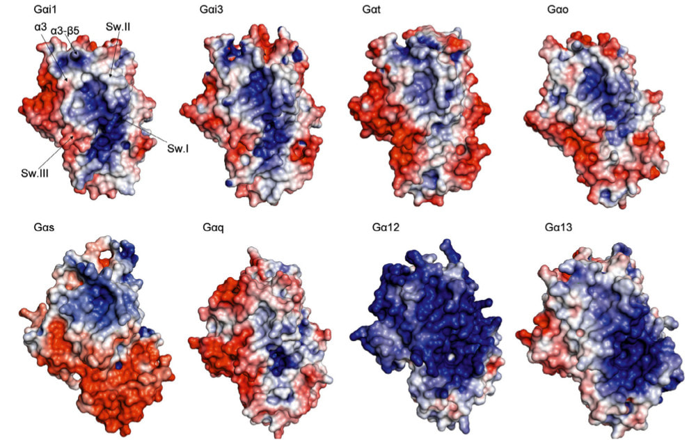
Molecular Dynamics Simulations
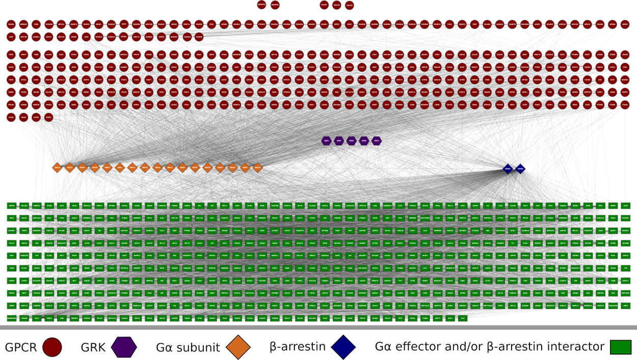
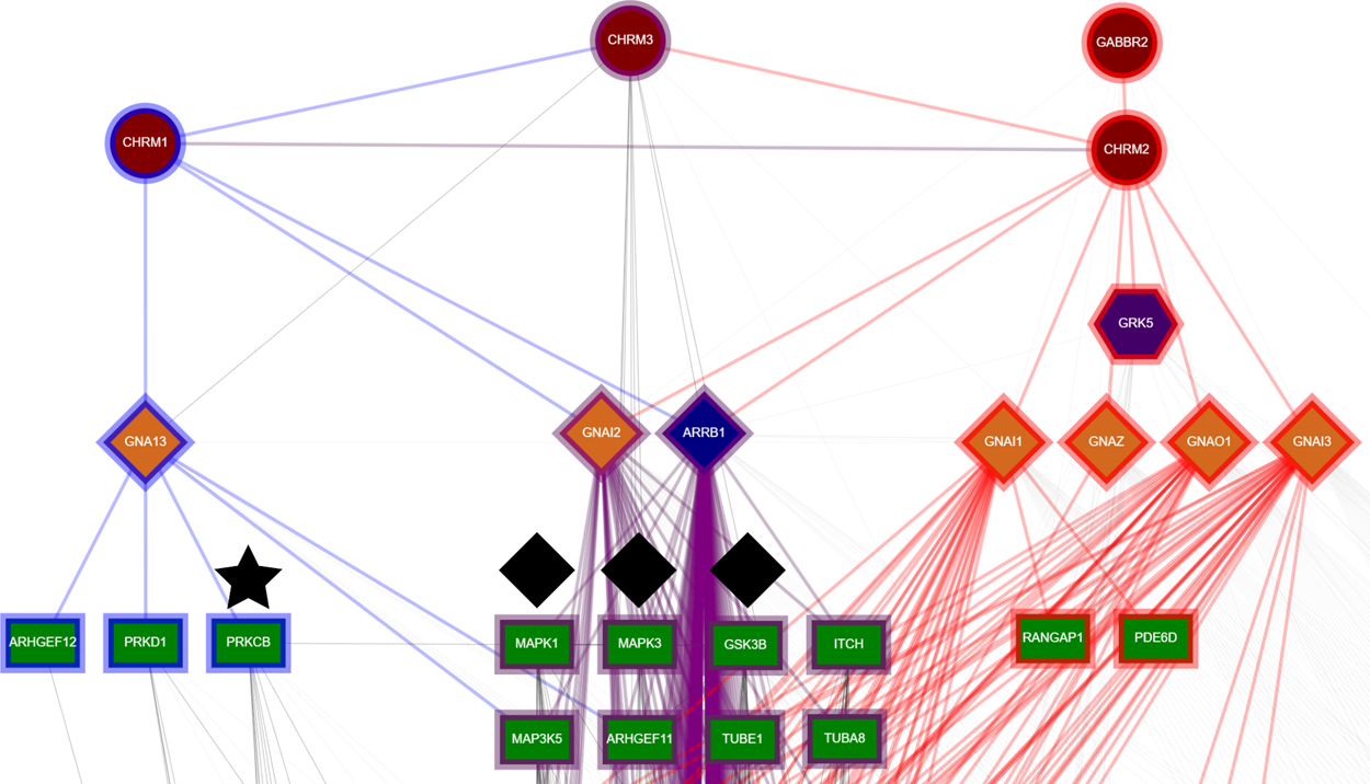
Molecular Dynamics simulations of LPS-containing outer membranes, resembling the OM of Escherichia coli (Source: Baltoumas et al, J. Comput. Chem. Jul 5; 40(18): 1727-1734, 2019).
Topology (A), three-dimensional structure (B-C) and protein-ion interactions (D-E) for the SLC11A1 divalent metal ion transporter (Source: Triantaphyllopoulos et al, J. Comput. Aid. Mol. Des. Feb; 33(2):265-285, 2019).
The proposed self-aggregation pathway for the formation of αCGRP amyloid fibrils, as indicated by Molecular Dynamics simulations combined with experimental assays (Source: Tsiolaki et al, J. Struct. Biol. Jul; 203(1):27-36, 2018).
Structural dynamics analysis of GPCR dimers through Dynamical Network Analysis, combining the results of MD simulations with Network Theory (Source: Baltoumas et al, J. Comput. Aided. Mol. Des. Jun;30(6):489-512, 2016).
Proposed model of the Pmel17 RPT domain in the acidic pH conditions of melanosomes (left), and its dissociation mechanism in neutral pH solvent (right) (Source: Louros et al, J. Comput. Aided. Mol. Des. Feb; 30 (2): 153-64, 2016).
Comparison of electrostatic potential distributions among different members of the Gα subunit superfamily (Source: Baltoumas et al, J. Struct. Biol. Jul; 182 (3), 209-218, 2013).

Biological Networks

The extended Human GPCR signaling network. The nodes have been laid out in a hierarchical manner, according to the flow of information in a cell. The top layer consists of GPCRs who receive the extracellular signal. At the upper most layers are found GPCRs that do not interact with any Gα subunits or β-arrestins. Following is the layer of GRKs that regulate GPCR binding to Gα subunits and β-arrestins via phosphorylation. In the middle layer lie the transducers, the main connectors of the network; GPCRs relay information to them and they, in turn, act by regulating an array of effector proteins, which make up the bottom layer of the network (Source: Apostolakou et al, J. Proteome Res. 19 (1), 511-524)
Comparison between the signaling pathways CHRM1 (blue) and CHRM2 (red). Common signaling pathways are displayed with the color purple. A diamond denotes the proteins MAPK3, MAPK1, and GSK3B that have a purple border because they are in both, CHRM1 and CHRM2, signaling pathways. A star denotes PRKCB, a PKC, part of CHRM1 signaling pathway and therefore with a blue border (Source: Apostolakou et al, J. Proteome Res. 19 (1), 511-524).

Journal Covers

Tsiolaki, P.L., Nasi, G.I., Baltoumas, F.A., Louros, N.N., Magafa, V., Hamodrakas, S.J. & Iconomidou, V.A. (2018) “αCGRP, another amyloidogenic member of the CGRP family” J. Struct. Biol. Jul; 203(1):27-36.
Louros, Ν.Ν., Tsiolaki, P.L., Baltoumas, F.A., Chryssikos, G.D., Gionis, V., Hamodrakas, S.J. & Iconomidou, V.A. (2017) "Tracking the amyloidogenic core of IAPP amyloid fibrils: Insights from micro-Raman spectroscopy". J. Struct. Biol. Aug; 199 (2): 140-152
Baltoumas, F.A., Theodoropoulou, M.C. & Hamodrakas, S.J. (2013) "Interactions of the α-subunits of heterotrimeric G-proteins with GPCRs, effectors and RGS proteins: a critical review and analysis of interacting surfaces, conformational shifts, structural diversity and electrostatic potentials". J. Struct. Biol. Jul; 182 (3), 209-218

Conference Posters


Conference Presentations

HeCrA 2018

Protein Summer School 2016

HSCBB 2015

HSCBB 2014


Videos

Powered by Create your own unique website with customizable templates.